Understanding Bladder Cancer
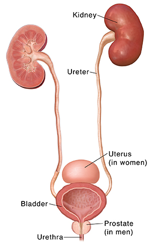
The urinary system includes the kidneys, ureters, bladder, and urethra. This body system makes, stores, and gets rid of liquid waste called urine. The two kidneys filter blood to collect waste and make urine. The two ureters are small tubes that carry urine from each kidney to the bladder. The bladder is where urine is stored before it leaves the body. The urethra is the tube that urine goes through to leave the body.
Bladder cancer means that cells in the bladder have changed in ways that aren't normal.
When bladder cancer forms
Cancer starts when cells change (mutate) and grow out of control. The dividing cells may form a lump of tissue (tumor ). With time, the cancer cells destroy healthy bladder tissue. They may spread to other parts of the body.
Why some cells become cancer is not always clear. But bladder cancer is strongly linked to cigarette smoking. In fact, smoking is linked to about half of all bladder cancers. The longer a person smokes and the more a person smokes, the greater that person’s chances of developing bladder cancer.
Types of cancer that may form
Most bladder cancers are urothelial carcinomas, also called transitional cell carcinomas. They start in the cells that make up the lining inside of the bladder. Bladder cancer may grow in different ways:
-
Papillary tumors. These stick out from the bladder lining on a stalk. They tend to grow toward the bladder cavity, away from the bladder wall, instead of deeper into the layers of the bladder wall.
-
Flat or sessile tumors. These don't stick out from the bladder lining. These tumors are much more likely than papillary tumors to grow into the deeper layers of the bladder wall.
-
Carcinoma in situ. This is a patch of flat tumor cancer cells that's only in the inner layer of the bladder lining and has not spread to deeper layers. The patch may look almost normal. Or it may look inflamed and red.
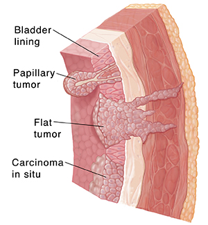
Each type of tumor can be found in more than one part of the bladder. And more than one type can form at the same time.
Online Medical Reviewer:
Anne Fetterman RN BSN
Online Medical Reviewer:
Raymond Turley Jr PA-C
Online Medical Reviewer:
Warren Brenn
Date Last Reviewed:
9/1/2025
© 2000-2026 The StayWell Company, LLC. All rights reserved. This information is not intended as a substitute for professional medical care. Always follow your healthcare professional's instructions.